
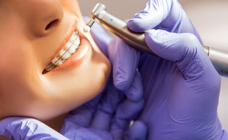
Radiographs or dental X-rays are one of the cornerstones of contemporary dental care. Although a visual dental examination will inform the dentist much about the well-being of your teeth and gums, it cannot reveal everything. X-rays enable clinicians to visualise what is occurring beneath the face, inside the teeth, under the gum, and within the jawbone, giving vital information required to diagnose, treat, and prevent dental issues before they escalate to serious consequences.
Why are Dental X-Rays Important?
Dental X-rays are mainly used to provide the dentist with a comprehensive perspective of your oral health, which could not have been reached with a clinical examination. They allow dentists to examine the areas that cannot be seen during a normal visual examination like between teeth and under existing restorations like fillings. This timely and thorough perception aids in the identification and resolution of problems at an earlier stage before they become more severe and invasive.
What Do Dental X-Rays Show?
Dental radiograph may expose any number of concerns that may not otherwise be apparent and include:
Minimal spots of caries amongst teeth that cannot be viewed through the eyes.
Beneath pre-existing fillings cavities or decay.
Around the roots of the teeth, there is an abscess or an infection.
Periodontal (gum) disease results in bone loss in the jaw.
Jaw or tissue cysts or tumours.
Arranging teeth in a way that helps in planning the treatment of implants, braces, dentures, or any other treatment of the teeth.
These lessons allow making a more accurate diagnosis and creating a treatment plan that is more personal.
Types of Dental X-Rays
There are various categories of dental radiographs and each one of them has various diagnostic purposes:
Bitewing X-rays: This method is mostly applied in the detection of early decay between back teeth.
Periapical X-rays: This is aimed at the roots of one or two teeth, useful in the examination of root health and the revealing of abscesses.
Panoramic X-rays: These are taken outside the mouth, and they give a complete view of the mouth, the teeth, jaws and joints.
Additional ones such as occlusal and cephalometric X-rays may help to make certain evaluations such as tracking the growth of teeth in children or orthodontic patient planning.
How Often Are X-Rays Needed?
Dental X-rays do not have any standardized schedule. The frequency at which you need them varies with your personal oral health, age, disease tendency and whether you have active disease problems. Others of good oral health may need to have radiographs after every couple of years, whereas those who have continuing problems may need to replace their radiographs after six months so as to keep up with changes. An initial patient will be offered a panoramic X-ray when the dentist wants to examine the oral state as a whole.
Are Dental X-Rays Safe?
The issue of safety is generally raised, however, the levels of radiation in dental X-rays are very low and with modern digital technology, these levels are even lower. The amount of radiation that a person receives per year due to dental imaging is a minor portion of the total dose received due to natural sources. Dentists comply with the safety measures by ensuring that their X-ray beam is based on the area of interest and protective shielding is used where necessary. Dental health authorities say that dental X-rays are healthy when applied in a responsible manner to both children and adults.


